Summary
The prevalence of subclavian, brachiocephalic and vertebral artery occlusive disease due to atherosclerotic disease in the general population is unknown as these conditions often remain undiagnosed because, very frequently, they are either asymptomatic or are due to neglected symptoms of vertebro-basilar and/or upper extremity ischaemia.
Subclavian and brachiocephalic endovascular interventions are procedures that are both feasible and acceptably safe. Nevertheless, the need for correct clinical judgment and patient selection must be emphasised.
In vertebral artery disease, asymptomatic subjects do not require revascularisation. In general, it is only patients with recurrent ischaemic events despite antiplatelet therapy or with refractory vertebro-basilar hypoperfusion in whom revascularisation should be considered.
With regard to subclavian artery disease, revascularisation is rarely indicated in asymptomatic patients [11. European Stroke Organisation; Authors/Task Force Members, Tendera M, Aboyans V, Bartelink ML, Baumgartner I, Clément D, Collet JP, Cremonesi A, De Carlo M, Erbel R, Fowkes FG, Heras M, Kownator S, Minar E, Ostergren J, Poldermans D, Riambau V, Roffi M, Röther J, Sievert H, van Sambeek M, Zeller T; ESC Committee for Practice Guidelines, Bax J, Auricchio A, Baumgartner H, Ceconi C, Dean V, Deaton C, Fagard R, Funck-Brentano C, Hasdai D, Hoes A, Knuuti J, Kolh P, McDonagh T, Moulin C, Poldermans D, Popescu B, Reiner Z, Sechtem U, Sirnes PA, Torbicki A, Vahanian A, Windecker S; Document Reviewers, Kolh P, Torbicki A, Agewall S, Blinc A, Bulvas M, Cosentino F, De Backer T, Gottsäter A, Gulba D, Guzik TJ, Jönsson B, Késmárky G, Kitsiou A, Kuczmik W, Larsen ML, Madaric J, Mas JL, McMurray JJ, Micari A, Mosseri M, Müller C, Naylor R, Norrving B, Oto O, Pasierski T, Plouin PF, Ribichini F, Ricco JB, Ruilope L, Schmid JP, Schwehr U, Sol BG, Sprynger M, Tiefenbacher C, Tsioufis C, Van Damme H. ESC Guidelines on the diagnosis and treatment of peripheral artery diseases: Document covering atherosclerotic disease of extracranial carotid and vertebral, mesenteric, renal, upper and lower extremity arteries: the Task Force on the Diagnosis and Treatment of Peripheral Artery Diseases of the European Society of Cardiology (ESC). Eur. Heart J. 2011;32:2851-906. ]. In symptomatic patients, even if endovascular and surgical treatment options are equally feasible, atherosclerotic lesions of the upper extremities, mostly subclavian lesions, are nowadays treated primarily by endovascular techniques.
As for carotid artery stenting, the importance of two major determinants for procedural success must be emphasised, namely, that these interventions should be based always on a pre-planned individual treatment strategy and performed by highly trained operators.
Subclavian, brachiocephalic and vertebral intervention
DEFINITION AND CLINICAL PRESENTATION
Subclavian artery disease is most frequently caused by atherosclerosis, and other etiologies include congenital malformations, fibro muscular dysplasia, neurofibromatosis, inflammation (e.g. Takaysu’s and other forms of arteritis), radiation exposure and mechanical causes (e.g. trauma or compression syndromes) [3535. Ochoa VM and Yeghiazarians Y. Subclavian artery stenosis: a review for the vascular medicine practitioner. Vascular medicine (London, England) 2011;16:29-34. , 3636. Bradaric C, Kuhs K, Groha P, Dommasch M, Langwieser N, Haller B, Ott I, Fusaro M, Theiss W, von Beckerath N, Kastrati A, Laugwitz KL, Ibrahim T. Endovascular therapy for steno-occlusive subclavian and innominate artery disease. Circ J. 2015;79:537-43. ].The subclavian arteries and the brachiocephalic trunk are the most common locations for atherosclerotic plaques in upper extremities. The left subclavian artery is more likely to be affected than the right. When thebrachiocephalic circulation is involved, the innominate artery is diseased in one third of cases, with the remaining two thirds affecting the right subclavian artery [3636. Bradaric C, Kuhs K, Groha P, Dommasch M, Langwieser N, Haller B, Ott I, Fusaro M, Theiss W, von Beckerath N, Kastrati A, Laugwitz KL, Ibrahim T. Endovascular therapy for steno-occlusive subclavian and innominate artery disease. Circ J. 2015;79:537-43. ].
Most commonly, a unilateral subclavian stenosis is detected following measurement of unequal arm pressures. Symptoms of subclavian or brachiocephalic trunk disease may include angina – in patients who have undergone coronary artery bypass surgery and have a stenosis proximal of an internal mammary artery graft – and vertebrobasilar insufficiency (VBI), such as subclavian steal syndrome (dizziness, vertigo, blurred vision, alternating hemiparesis, dysphasia, dysarthria, confusion and loss of consciousness), drop attacks, ataxia or other postural disturbances including sensory and visual changes. Ischaemic arm symptoms are usually of minor clinical relevance. Patients with previous axillofemoral bypass may develop intermittent claudication. Brachiocephalic occlusive disease may also cause ischaemic neurological symptoms.
Knowing how to properly treat a diseased subclavian artery is then far relevant for all interventional cardiologists dealing with patients who previously received surgical revascularization or presenting with concomitant peripheral arteriopathy.
IMAGING
The proximal location of subclavian and brachiocephalic occlusive disease makes duplex ultrasonography a challenge. However, duplex scanning is of particular value in differentiating occlusion from stenosis, in determining the direction of vertebral blood flow and in screening for concurrent carotid artery stenosis. Nowadays, CT angiography offers excellent anatomic resolution with precise information on lesion morphology, length and location. However, it does not provide optimal information on the degree of calcification [3737. Dosa E, Nemes B, Berczi V, Novak PK, Paukovits TM, Sarkadi H and Huttl
K. High frequency of brachiocephalic trunk stent fractures does not impair clinical outcome. Journal of vascular surgery. 2014;59:781-5. ]. On the other hand, MR angiography can be misleading as reduced flow can be interpreted as exaggerated disease [3838. Ibrahim T. How should I treat a complex left subclavian artery stenosis
involving the vertebral artery in a patient with subclavian steal syndrome and left internal mammary artery bypass graft? . EuroIntervention. 2015;10:e1-e7. ]. Therefore, digital subtraction angiography – still considered as the imaging “gold standard” – is rarely required for diagnostic purposes.
REVASCULARISATION OPTIONS
Expert opinion favors an endovascular strategy as first line therapy. Compared to angioplasty, stenting has a superior patency rate and avoids recoil and restenosis [3939. Chatterjee S, Nerella N, Chakravarty S and Shani J. Angioplasty alone versus angioplasty and stenting for subclavian artery stenosis--a systematic review and meta-analysis American journal of therapeutics. 2013;20:520-3. ]. Endovascular interventions have a success rate of approximately 95% with a 2.3-3.6% combined risk of stroke and death [4040. De Vries JP, Jager LC, Van den Berg JC, Overtoom TT, Ackerstaff RG, Van de Pavoordt ED and Moll FL. Durability of percutaneous transluminal angioplasty for obstructive lesions of proximal subclavian artery: long-term results. Journal of vascular surgery. 2005;41:19-23. ]. Primary and secondary patency rates at ten years are 78.1% and 84.5% respectively [4141. Henry M, Henry I, Polydorou A, Polydorou A and Hugel M. Percutaneous transluminal angioplasty of the subclavian arteries. International angiology : a journal of the International Union of Angiology. 2007;26:324-40. ].
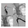
From a technical standpoint, the primary anatomical consideration when stenting the proximal left subclavian artery is the location of the left vertebral artery. Appropriate angiographic angulations should be acquired to see the true ostium of the vertebral artery prior to stent implantation. The majority of cases will allow a landing zone prior to the vertebral origin ( Figure 1 ). When there is no landing zone prior to the vertebral artery, the risk of the procedure may increase due to possible plaque shift into the vertebral artery. However, a recent paper by Schneider et al. Showed that endovascular revascularization of subclavian artery disease with medial segments involving the vertebral artery origin required more complex techniques but showed long-term patency rates comparable to those in lesions located within the proximal vessel [4242. Schneider V, Dirschinger R, Wustrow I, Müller A, Cassese S, Fusaro M, Kastrati A, Koppara T, Bergmann K, Laugwitz KL, Ibrahim T, Bradaric C. Endovascular therapy of subclavian artery occlusive disease involving the vertebral artery origin. Vasa 2020;49:205-213. ]. When approaching the right system the operator must adequately visualise the bifurcation of the right common carotid and right vertebral arteries when repairing the innominate and right subclavian arteries, respectively. Typically the bifurcation of the right common carotid and innominate artery is best seen in the RAO projection. In the setting of significant atherosclerotic burden distal protection within the vertebral system has been advocated by some, although the risk of stroke is small, potentially due to delayed reversal of vertebral artery blood flow from retrograde to antegrade following angioplasty of the subclavian artery [22. Nasim A, Sayers RD, Bell PR, Bolia A. Protection against vertebral artery embolisation during proximal subclavian artery angioplasty. Eur J Vasc Surg. 1994;8: 362-3.
Technical aspects of proximal subclavian artery angioplasty, 33. Ringelstein EB, Zeumer H. Delayed reversal of vertebral artery blood flow following percutaneous transluminal angioplasty for subclavian steal syndrome. Neuroradiology. 1984;26:189-98. ]. Likewise, distal protection within the right internal carotid artery during repair of the right innominate artery may be considered for bulky atherosclerotic lesions.
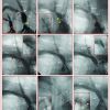
Access for endovascular repair of the innominate or subclavian artery can be via the common femoral (most frequently), brachial, or radial arteries. The femoral approach will permit the use of either a sheath or a guide catheter. When using a sheath, the operator can either wire the lesion through a diagnostic catheter and replace the entire system with a sheath (usually 6-7 Fr and 90 cm in length) or more simply telescope the diagnostic catheter through the sheath and once wired advance the sheath into the appropriate location over the diagnostic catheter. Guiding catheters are limited by their smaller internal diameter when compared to that of similar sized sheaths. Sheaths are routinely used during brachial (6-7 Fr, 45 cm) and radial (6 Fr, 65-90cm) access. Once the lesion has successfully been crossed with a standard 0.035” wire, balloon predilatation can be used to gauge size and length prior to stent implantation. Subclavian arteries usually accommodate 7-10 mm diameter stents while innominate arteries may require larger devices. Balloon-expandable stents are primarily used for their radial strength and precise placement. If the lesion is located aorto-ostially, the stent should protrude 1-2mm into the aorta in order to have a complete coverage of the lesion. Although most investigators believe that no distal protection of the vertebral artery is required at the time of proximal upper extremity intervention careful consideration must be given to protecting the right internal carotid for bulky innominate lesions. Protection of the right internal carotid can be accomplished via access and deployment of the protection device through an additional sheath in the right brachial or radial artery while primarily working through a sheath from the femoral to perform the stenting procedure ( Figure 2 ).
When to intervene in patients with subclavian and brachiocephalic disease
- Revascularisation is indicated only in symptomatic patients
- In patients with atherosclerotic lesions of brachiocephalic trunks, endovascular approach is indicated as first treatment strategy
- Revascularisation of an asymptomatic patient with severe stenosis of brachiocephalic trunks may be indicated only in case of former or future mammary-coronary bypass, or for monitoring blood pressure in bilateral upper limb occlusion
Extracranial vertebral artery interventions
DEFINITION AND CLINICAL PRESENTATION
The prevalence of atherosclerotic occlusive disease of the extracranial vertebral artery (VA) in the general population is unknown because this condition often remains undiagnosed, either because asymptomatic or because the symptoms of vertebrobasilar ischaemia are not recognised. It has been estimated that approximately 20% of all ischaemic strokes involve the vertebrobasilar territory [44. Bogousslavsky J, Van Melle G, Regli F. The Lausanne Stroke Registry: analysis of 1,000 consecutive patients with first stroke. Stroke; a journal of cerebral circulation. 1988;19:1083-92. , 55. Bogousslavsky J, Regli F. Borderzone infarctions distal to internal carotid artery occlusion: prognostic implications. Ann Neurol. 1986;20:346-50. ]. Although it was long believed that the risk of recurrent stroke was lower for the posterior compared with the anterior circulation, a meta-analysis has suggested that overall the two vascular territories carry a similar risk of recurrence [66. Flossmann E, Rothwell PM. Prognosis of vertebrobasilar transient ischaemic attack and minor stroke. Brain. 2003;126:1940-54. ]. With regard to the underlying pathology, it is estimated that a haemodynamic significant (≥50%) stenosis of the extracranial VA may account for 1/5 of all vertebrobasilar stroke or TIA [77. Caplan LR, Wityk RJ, Glass TA, Tapia J, Pazdera L, Chang HM, Teal P, Dashe JF, Chaves CJ, Breen JC, Vemmos K, Amarenco P, Tettenborn B, Leary M, Estol C, Dewitt LD, Pessin MS. New England Medical Center Posterior Circulation registry. Ann Neurol. 2004;56:389-98. ].
Vertebrobasilar ischaemia is mostly the result of an embolic process – most frequently as artery-to-artery embolism from the VA origin or alternatively as a cardio-embolic process; on occasion thrombotic and low-flow haemodynamic mechanisms may play a role [88. Wityk RJ, Chang HM, Rosengart A, Han WC, DeWitt LD, Pessin MS, Caplan LR. Proximal extracranial vertebral artery disease in the New England Medical Center Posterior Circulation Registry. Arch Neurol. 1998;55:470-8. ]. Although atherosclerotic stenosis can occur at any point along the length of the VA, the most frequent location is at or near its origin [99. Schwartz CJ, Mitchell JR. Atheroma of the carotid and vertebral arterial systems. Br Med J. 1961;2:1057-63. ]. Atherosclerotic VA lesions have traditionally been believed to be benign based on the capacity to develop collateral reconstitution of the extracranial VA, the usual presence of two viable arteries that join together intracranially, and the slow development of luminal compromise by atherosclerotic plaques allowing time for collateral development [1010. Fisher CM. Occlusion of the vertebral arteries. Causing transient basilar symptoms. Arch Neurol. 1970;22:13-9. ].
IMAGING
Few high-quality studies are available on imaging of the extracranial vertebral artery and no studies have compared different imaging modalities against the gold standard contrast angiography in the same cohort of patients. It has been suggested that magnetic resonance angiography (MRA) offers better sensitivity and specificity than duplex ultrasonography for extracranial VA stenosis [1111. Khan S, Cloud GC, Kerry S, Markus HS. Imaging of vertebral artery stenosis: a systematic review. J Neurol Neurosurg Psychiatry. 2007;78:1218-25.
Role of imaging in vertebral artery lesion assessment]. However, due to the high variability of flow in different anatomical subsets, it could overestimate the lesions. Computed tomography angiography (CTA) is increasingly being used as the imaging modality of choice to replace contrast angiography in many centres for extracranial VA disease [1111. Khan S, Cloud GC, Kerry S, Markus HS. Imaging of vertebral artery stenosis: a systematic review. J Neurol Neurosurg Psychiatry. 2007;78:1218-25.
Role of imaging in vertebral artery lesion assessment]. However, VIST highlighted difficulties in diagnosis of vertebral stenosis in clinical practice. Although previous studies have shown good sensitivity and specificity of computed tomographic angiography and contrast enhanced magnetic resonance angiographic in detection of vertebral stenosis compared with intra-arterial digital subtraction angiography (DSA) [4343. Khan S, Rich P, Clifton A, Markus HS. Noninvasive detection of vertebral artery stenosis: a comparison of contrast-enhanced MR angiography, CT angiography, and ultrasound. Stroke 2009; 40:3499–3503. ], in VIST, in 23 of 91 patients randomized to stenting, stenosis of more than 50% was not confirmed on DSA performed at the time of planned stenting [4444. Markus HS, Larsson SC, Kuker W; VIST Investigators. Stenting for symptomatic vertebral artery stenosis: the Vertebral Artery Ischaemia Stenting Trial. Neurology 2017; 89:1229–1236. ]. Central review of entry imaging failed to confirm stenosis in approximately half of the cases that had been deemed to have stenosis by the recruiting centre, whereas in others, entry imaging was adjudged to be of insufficient quality to confirm stenosis.
From a practical standpoint, both MRA and CTA may be inadequate for ostial VA lesions, especially in the presence of severe angulation or tortuosity of the VA take-off. In these cases, if a severe stenosis needs to be excluded with certainty, contrast angiography remains the diagnostic tool of choice.
MANAGEMENT
Atherosclerotic extracranial VA occlusive disease may be approached medically, using endovascular techniques, or surgically. While the optimal antithrombotic therapy – aspirin, clopidogrel, or warfarin – in VA stenosis has not been defined, most patients are treated with low-dose aspirin.
Various types of surgical interventions include vertebral artery repair, vertebral artery transposition, and vertebral artery bypass. However, the number of expert surgeons able to perform such kind of surgery is limited, so endovascular techniques are now preferred.
With respect to endovascular revascularisation, the risk of the intervention must be outweighed by the potential benefits of revascularisation. The need to intervene is tempered by the fact that the posterior circulation is supplied by the confluence of the two VA and a large proportion of patients remain asymptomatic despite occlusion of one extracranial VA. Although surgery of extracranial VA stenosis has been performed with low stroke and mortality rates by surgeons with extensive experience [1212. Berguer R, Flynn LM, Kline RA, Caplan L. Surgical reconstruction of the extracranial vertebral artery: management and outcome. J Vasc Surg. 2000;31:9-18. ], in most centres the surgical approach is not considered a viable alternative to endovascular treatment because it is far more invasive.
Patients with symptoms attributed to vertebrobasilar ischaemia and isolated stenosis of the extracranial VA >50% confirmed on angiography may benefit from revascularisation, especially if the vessel is dominant or the contralateral site occluded. The majority of patients with asymptomatic extracranial VAdisease do not require treatment. In asymptomatic patients with high-grade (>70%) stenosis revascularisation may be considered, especially if the severity of the lesion is progressive and the affected VA is dominant or is the only patent one.
TECHNICAL ASPECTS OF VERTEBRAL INTERVENTIONS
Most vertebral interventions can be performed with coronary equipment using a 6 Fr guiding catheter, a medium or extra support 0.014” coronary guidewire and a balloon-expandable coronary or peripheral stent [1313. Mukherjee D, Roffi M, Kapadia SR, Bhatt DL, Bajzer C, Ziada KM, Kalahasti V, Hughes K, Yadav JS. Percutaneous intervention for symptomatic vertebral artery stenosis using coronary stents. J Invasive Cardiol. 2001;13:363-6. ]. Guiding catheter shapes frequently used include the Judkins right or vertebral shapes. Since the lesions are frequently in ostial locations, coronary stents with good radial strength are preferred. Although formal comparisons are missing, stenting has virtually replaced angioplasty in the treatment of extracranial VA stenosis.
Similar to CAS, endovascular treatment of extracranial VA stenosis is usually performed under local anaesthesia and conscious sedation, allowing for early detection of neurological symptoms. After arterial access is obtained, usually using a femoral approach, unfractionated heparin is administered to achieve full anticoagulation. On occasion, the radial or brachial artery access may be preferred because of unfavourable anatomy at the level of the aortic arch or excessive angulation between the subclavian artery and the origin of the VA. A 6 Fr guide catheter is advanced over a 0.035” wire to obtain a stable position in the subclavian artery. Usual curves include the vertebral, Judkins right, and the multipurpose one. If additional stability is needed, the use of a 7 Fr guide catheter or 7 Fr 90 cm sheath may be helpful because it allows the advancement of a 0.014” or 0.018” “buddy” wire into the distal subclavian artery. If the long sheath approach is chosen, then a 0.035”exchange length wire is advanced over a diagnostic catheter positioned in the SCA in the axillary artery and then the long sheath is advanced over the wire. Angiographic runs are performed to visualise the extracranial and intracranial VA and to obtain accurate views defining the VA lesions and their relation to the subclavian artery.
Although embolisation may occur during vertebral interventions, the use of EPD is controversial since it may be associated with complications such as dissection or spasm [1414. Canyigit M, Arat A, Cil BE, Turkbey B, Saatci I, Cekirge S, Balkanci F. Distal embolization after stenting of the vertebral artery: diffusion-weighted magnetic resonance imaging findings. Cardiovasc Intervent Radiol. 2007;30:189-95.
Role of diffusion-weighted magnetic resonance imaging for assessing complications after stenting of the vertebral artery]. Accordingly, the smaller calibre of the VA, the frequently marked angulation between the VA origin and the subclavian artery, the tortuosity of the proximal VA segments, and the tendency to develop spasms are all factors that may cause difficulties in the advancement of the EPD. In addition, recovery of the system may also be difficult in the presence of spasms or unfavourable angulation of the VA origin, angulation that may be more pronounced following stent deployment. Therefore, embolic protection devices are usually not recommended for VA interventions. On occasion, the use ofEPD may be considered for VA interventions for lesions at high embolic risk in the presence of VA with large diameter, favourable geometric orientation of the VA origin, and absence of significant tortuosity.
Under road-map guidance, the VA lesion is crossed with a 0.014” medium-support or extra-support coronary wire. In order to obtain a stable guiding catheter position, the wire should be positioned far enough distally in the VA. The tip of the wire should be visualised during the entire procedure to reduce the risk of artery perforation. Stent selection (either self-expanding or balloon-mounted) is based mainly on lesion location. Ostial VA lesions are treated preferably with balloon-expandable coronary stents because of high radial force, lack of foreshortening and low crossing profile. For true ostial lesions, the stent should protrude 1-2 mm into the SCA to allow for optimal lesion coverage and a second balloon inflation at high pressure should be performed following partial balloon retrieval to optimise the apposition of the stent struts at the vessel wall, the so-called “flaring” of the ostium. Self-expanding stents are reserved for non-ostial lesions in vessels with larger diameters (i.e., >5.5 mm) [1515. Wehman JC, Hanel RA, Guidot CA, Guterman LR, Hopkins LN. Atherosclerotic occlusive extracranial vertebral artery disease: indications for intervention, endovascular techniques, short-term and long-term results. J Interv Cardiol. 2004;17:219-32.
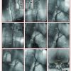
Patient selection and indication to treatment of occlusive extracranial vertebral artery disease]. In the presence of a severe or calcified lesion, balloon dilatation prior to stenting may be appropriate. The balloon selected should be undersized and shorter than the stent placed thereafter ( Figure 2 and Figure 3 ).
DATA ON VERTEBRAL INTERVENTIONS
A randomised controlled trial (Carotid And Vertebral Artery Transluminal Angioplasty Study – CAVATAS) compared angioplasty and stenting (n=8) to medical therapy (n=8) for symptomatic VA proximal artery stenosis [1616. Coward LJ, McCabe DJ, Ederle J, Featherstone RL, Clifton A, Brown MM; CAVATAS Investigators. Long-term outcome after angioplasty and stenting for symptomatic vertebral artery stenosis compared with medical treatment in the Carotid And Vertebral Artery Transluminal Angioplasty Study (CAVATAS): a randomized trial. Stroke. 2007;38:1526-30. ]. The trial showed no benefit of endovascular treatment over medical regime. No patient in either arm experienced a recurrent stroke in the vertebrobasilar territory up to 4,7 years after randomisation. Criticism of this trial has been made due to the small number (n=16) of patients involved.
The Stenting of Symptomatic Atherosclerotic Lesions in the Vertebral or Intracranial Arteries (SSYLVIA) study is a prospective multi-centre study addressing the value of a dedicated stent for VA interventions [1717. SSYLVIA Study Investigators. Stenting of Symptomatic Atherosclerotic Lesions in the Vertebral or Intracranial Arteries (SSYLVIA): study results. Stroke. 2004;35: 1388-92.
The best prospective multicentre study evaluating a dedicated stent for vertebral interventions]. However, among the 61 patients enrolled, 18 also included the extracranial VA. In this subgroup, two patients suffered a stroke at 1 year (11%). Six of 14 patients (43%) who underwent repeat angiography at six months had a restenosis >50%.
In the same years, a large series including 112 consecutive procedures in 105 symptomatic patients over a 12-year period reported a technical success of 100%. Periprocedural complications included one flow-limiting dissection, two bleedings requiring transfusion, one access site complication and one TIA (4.8% per patient) [1818. Jenkins JS, Patel SN, White CJ, Collins TJ, Reilly JP, McMullan PW, Grise MA, Grant AG, Ramee SR. Endovascular stenting for vertebral artery stenosis. J Am Coll Cardiol. 2010;55:538-42.
A balanced overview of vertebral artery stenting techniques]. No patient suffered periprocedural stroke. Vertebrobasilar stroke occurred in two patients at 30 days and in addition three patients at one year (total stroke rate at one year 4.8%). While the restenosis rate is not reported, the target vessel revascularisation rate and sustained symptom resolution at a median follow-up of 29 months were 13% and 70%, respectively.
Two more recent trials (VIST and VAST: Vertebral Artery Stenting Trial) [4444. Markus HS, Larsson SC, Kuker W; VIST Investigators. Stenting for symptomatic vertebral artery stenosis: the Vertebral Artery Ischaemia Stenting Trial. Neurology 2017; 89:1229–1236. , 4545. Compter A, van der Worp HB, Schonewille WJ, Vos JA, Boiten J, Nederkoorn PJ, Uyttenboogaart M, Lo RT, Algra A, Kappelle LJ; VAST investigators. Stenting versus medical treatment in patients with symptomatic vertebral artery stenosis: a randomised open-label phase 2 trial. Lancet Neurol 2015; 14:606–614. ] included both extracranial and intracranial stenoses. VAST and VIST recruited patients with symptomatic vertebral extracranial and intracranial artery stenosis and used more modern interventional equipment and predominantly stenting.
VAST aimed for a target of 180 patients but only recruited 115 (83% of which had extracranial stenosis) due to early closure secondary to regulatory issues. A total of 57 patients were assigned to stenting and 58 to medical treatment alone with median interval between last symptoms and randomization of 25 days (IQR 11–50). During a median follow-up of 3 years (IQR 1.3–4.1), there were eight strokes in the stenting group and seven in the medical treatment group. More early strokes (within 30 days) occurred in the stenting arm (three vs. one), and more strokes occurred during long-term follow-up in the medical arm (seven vs. four). Of the three early strokes in the stenting arm, two occurred in the nine patients with intracranial stenosis (22%) and only one in the 48 (2%) patients with extracranial stenosis. Although VAST was underpowered to determine whether stenting was more effective than medical treatment, the overall results show no difference in outcome. They do, however, confirm a low perioperative risk for extracanial stenosis, but much higher risk for intracranial stenosis.
VIST aimed to recruit 540 patients with at least 50% symptomatic vertebral stenosis, but recruitment was closed by the funder after 181 patients were enrolled due recruitment targets not being met. Three patients did not contribute follow-up data, leaving 91 patients in the stent group and 88 in the medical group. Mean follow-up was 3.5 years (IQR 2.1–4.7). Of 61 stented patients, stenosis was extracranial in 48 (78.7%) and intracranial in 13 (21.3%). No periprocedural complications occurred with extracranial stenting; two strokes occurred during intracranial stenting. The primary endpoint of fatal or nonfatal stroke occurred in five patients in the stent group vs. 12 in the medical group [hazard ratio 0.40; 95% confidence interval (CI) 0.14–1.13, P=0.08], with an absolute risk reduction of 25 strokes per 1000 person-years.
The time from last symptoms to randomization was shorter in the stenting arm by mean 12.8 days (P=0.04), suggesting they might have been a higher risk group. When days from last symptoms were adjusted for, the corresponding hazard ratio for the primary endpoint was 0.34 (95% CI 0.12–0.98; P=0.046), suggesting a benefit in favour of stenting. When a second post-hoc analysis in patients randomized within 2 weeks after the last symptom was performed, the hazard ratio of the primary endpoint was 0.30 (95% CI 0.09–0.99; P¼0.048). Strengths of VIST include the randomized design, that it is the largest study of stenting for vertebral stenosis, and that no patients were lost to follow-up.
All larger studies about vertebral interventions included both intracranial and extracranial lesions. The higher risk of early recurrent stroke after stroke or TIA associated with intracranial, compared with extracranial, vertebral stenosis suggests that stenting might be particularly beneficial for intracranial stenosis. On the other hand, the higher rate of interventional complications reported for intracranial stenosis might suggest the opposite. Therefore, it is important that analyses are performed not only for all cases of vertebral stenosis, but also stratified by location. A recent preplanned pooled individual patient data analysis of the three major randomised controlled trials comparing stenting with medical treatment in patients with symptomatic vertebral stenosis [4646. Markus HS, Harshfield EL, Compter A, Kuker W, Kappelle LJ, Clifton A, van der Worp HB, Rothwell P, Algra A; Vertebral Stenosis Trialists' Collaboration. Stenting for symptomatic vertebral artery stenosis: a preplanned pooled individual patient data analysis. Lancet Neurol 2019;18:666-673. ].
Most relevant sizeable (n≥25) series of vertebral interventions are listed in Table 1 . Overall, the technical success is greater than 95% and periprocedural stroke is a rare event, being absent in most of the series and occurring in <5% of all of them. Restenosis appears to be the Achilles’ heel of the procedure. Its true incidence is unknown because the follow-up of most series was not systematic and the reported results varied considerably (between 3% and 52%). Although the placement of drug-eluting stents has proven to be highly efficacious in restenosis prevention in the coronary artery, the results in the vertebral artery are still sparse. Two series with more than 25 patients undergoing drug-eluting stent implantation in the VA have reported promising low restenosis rates of 7% and 12% [1919. Gupta R, Al-Ali F, Thomas AJ, Horowitz MB, Barrow T, Vora NA, Uchino K, Hammer MD, Wechsler LR, Jovin TG. Safety, feasibility, and short-term follow-up of drug-eluting stent placement in the intracranial and extracranial circulation. Stroke; a journal of cerebral circulation. 2006;37:2562-6. , 2020. Vajda Z, Miloslavski E, Güthe T, Fischer S, Albes G, Heuschmid A, Henkes H. Treatment of stenoses of vertebral artery origin using short drug-eluting coronary stents: improved follow-up results. AJNR Am J Neuroradiol. 2009;30:1653-6.
Role of drug eluting stents in vertebral artery stenting]. Similarly, a retrospective single-centre study of patients treated for ostial VA stenosis suggests a lower rate of restenosis (2/12; 17%) in patients with drug-eluting stents compared with bare metal stents (9/24; 38%) [2121. Ogilvy CS, Yang X, Natarajan SK, Hauck EF, Sun L, Lewis-Mason L, Hopkins LN, Siddiqui AH, Levy EI. Restenosis rates following vertebral artery origin stenting: does stent type make a difference? J Invasive Cardiol. 2010;22:119-24.
Restenosis after endovascular treatment of vertebral artery disease].
A meta-analysis published in 2014 showed that the use of DES was associated with significantly lower (p<0.0001) overall restenosis rates [8.2% (14/170) over follow-up periods averaging 16 to 43 months] compared to BMS [23.7% (68/287) over mean follow-up periods of 19 to 46 months]. Moreover, DES showed significantly lower symptomatic restenosis rates as compared to BMS [4.7% (8/169) for DES vs. 11.6% (32/275) for BMS; p=0.005] [4747. Langwieser N, Buyer D, Schuster T, Haller B, Laugwitz KL, Ibrahim T. Bare metal vs. drug-eluting stents for extracranial vertebral artery disease: a meta-analysis of nonrandomized comparative studies. J Endovasc Ther 2014;21:683-92. ].
When to intervene in patients with extracranial vertebral artery disease
- In patients with symptomatic extracranial vertebral artery stenosis, endovascular treatment may be considered for lesions ≥50% in the case of recurrent ischaemic events despite optimal medical therapy
- The use of an EPD should be considered on a case-by-case approach after evaluation of anatomical challenges and global embolic risk
- Revascularisation of an asymptomatic vertebral artery stenosis is not indicated, irrespective of the degree of stenosis


