Summary
In the past, the most frequent indication for performing EMB was to monitor the presence or absence of cardiac allograft rejection in patients who underwent cardiac transplantation. Today, the spectrum of possible EMB indications has broadened and comprises work-up of non-ischaemic cardiomyopathies such as viral myocarditis, inflammatory dilated cardiomyopathy (iDCM), infiltrative cardiomyopathies, hypertrophic cardiomyopathy (HCM) or restrictive cardiomyopathy, and rare myocardial diseases with potential life-saving therapeutic options such as giant cell myocarditis, cardiac amyloidosis or cardiac sarcoidosis. In principal, the potential benefits of the EMB information (the guidance of therapeutic decisions and prognosis) have to be weighed-up against pre-existing information based on non-invasive methods, and the risk of complications in each individual when performing invasive EMB.
Since numerous recent studies have shown that firstly, the information given by EMB work-up is of diagnostic, therapeutic and prognostic value and secondly, the complication risk of EMB is rather low when performed by experienced physicians, EMB is still the gold-standard for the diagnosis of most cardiomyopathies. However, a thorough knowledge regarding the correct indication for performing EMB, the practical details of the EMB procedure, important safety issues and the essential post-procedural EMB analysis tools are a prerequisite for a clinically meaningful and practically successful procedure. Hence, the following sections aim at illustrating clearly the essential aspects of performing EMBs.
A short history of endomyocardial biopsy
The first human heart biopsies were obtained by Sutton in 1956 through a limited thoracotomy using a Vim-Silverman biopsy needle [11. Sutton DC, Sutton GC. Needle biopsy of the human ventricular myocardium: review of 54 consecutive cases. Am Heart J. 1960;60:364-70. ]. However, owing to frequent complications such as cardiac tamponade and pneumothorax, this and other invasive approaches did not become popular. In 1962, the first percutaneous bioptome was developed and clinically used via the basilic or saphenous vein by Konno and Sakakibara in Japan and called consequently the Konno-Sakakibara bioptome (jaw diameter 2.5 mm and 3.5 mm, respectively) [22. Sakakibara S, Konno S. Endomyocardial biopsy. Jpn Heart J. 1962;3:537-43. ]. A few years later, this design was further simplified by Caves and Schulz in Stanford enabling biopsy sampling from both the right ventricular septum through the internal jugular vein (using a shorter and thicker bioptome) and the left ventricular wall (using a longer and thinner bioptome), and therefore named the Caves-Schulz-Stanford bioptome (jaw diameter 2.5 mm) [33. Caves PK, Schulz WP, Dong E Jr, Stinson EB, Shumway NE. New instrument for transvenous cardiac biopsy. Am J Cardiol. 1974;33:264-7. ]. This disposable instrument became very popular for the assessment of cardiac allograft rejection in North America and consequently, right ventricular biopsy was increasingly performed. However, the Konno-Sakakibara bioptome as well as the initial Caves-Schulz-Stanford bioptome were stiff devices that had to be maneuvered independently through the vasculature. Today, modified, more flexible devices that are positioned with the aid of a long sheath or introducing catheter are more commonly used (see section “Choice of bioptome”).
Current indications for endomyocardial biopsy
a) The primary goals of performing EMBs areto find the underlying diagnosis in a patient with ongoing symptoms of heart failure,
b) to be able to decide whether a specific and aggressive therapy (such as immediate immunosuppression in giant-cell myocarditis as well as cardiac sarcoidosis or targeted therapy in specific amyloidosis subforms) is required or should be continued,
c) to obtain valuable data regarding future risk stratification.
In a previous landmark study, Ardehali et al analysed 845 patients with unexplained cardiomyopathy who underwent EMB to evaluate how often this procedure changed the clinically established diagnoses in such patients [44. Ardehali H, Qasim A, Cappola T, Howard D, Hruban R, Hare JM, Baughman KL, Kasper EK. Endomyocardial biopsy plays a role in diagnosing patients with unexplained cardiomyopathy. Am Heart J. 2004;147:919-23.
Comprehensive retrospective study demonstrating the clinical value of endomyocardial biopsy information in patients with non-ischaemic cardiomyopathy.]. Their findings suggest that the initial clinical assessment of the aetiology of new-onset cardiomyopathy is inaccurate in about one third of patients who have unexplained cardiomyopathy.
In the past, there were ongoing doubts regarding the correct indication and the clinical as well as the therapeutic value of the invasive EMB procedure which has a non-negligible risk of complications. In particular, the disappointing results of the large multicentre Myocarditis Treatment Trial [55. Mason JW, O’Connell JB, Herskowitz A, Rose NR, McManus BM, Billingham ME, Moon TE. A clinical trial of immunosuppressive therapy for myocarditis. The Myocarditis Treatment Trial Investigators. N Engl J Med. 1995;333:269-75. ] which was performed in 31 centres located in the United States, Canada and Japan in the 1990’s damaged the popularity of EMB (especially in the United States). However, the diagnosis of myocarditis in that trial was based on the Dallas criteria (where immunohistochemistry was not applied) that are outdated today. Immunohistochemical and molecular pathological methods (as discussed in the section “Required analyses of EMB samples”) were shown to be more sensitive for the diagnosis of myocardial inflammation even in the absence of focal lymphocytic infiltration and have been used successfully to identify patients responding to immunomodulatory therapy independent of the Dallas criteria [77. Frustaci A, Chimenti C, Calabrese F Verardo R, Thiene G, Russo MA, Maseri A, Frustaci A. Immunosuppressive therapy for active lymphocytic myocarditis: virological and immunologic profile of responders versus nonresponders. Circulation. 2003;107:857-63. , 88. Wojnicz R, Nowalany-Kozielska E, Wojciechowska C Glanowska G, Wilczewski P, Niklewski T, Zembala M, Polonski L, Rozek MM, Wodniecki J. Randomized, placebo-controlled study for immunosuppressive treatment of inflammatory dilated cardiomyopathy: two-year follow-up results. Circulation. 2001;104:39-45. ].
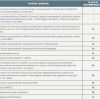
The recommendations of the American Heart Association (AHA)/American College of Cardiology (ACC)/European Societey of Cardiology (ESC), already published in 2007, comprised 14 different clinical scenarios in which performing EMB was deemed appropriate ( Table 1 ) [99. Cooper LT, Baughman KL, Feldman AM, Frustaci A, Jessup M, Kuhl U, Levine GN, Narula J, Starling RC, Towbin J, Virmani R. The role of endomyocardial biopsy in the management of cardiovascular disease: a scientific statement from the American Heart Association, the American College of Cardiology, and the European Society of Cardiology. Endorsed by the Heart Failure Society of America and the Heart Failure Association of the European Society of Cardiology. Eur Heart J. 2007;28:3076-93.
Clear scientific statements regarding the appropriate indications for endomyocardial biopsy.]. In principle, these recommendations are still valid and simplify appropriate clinical decision making, but cannot, of course, replace individual clinical assessment. In view of the perceived high risk of performing EMB, the 2007 recommendations were rather restrictive in defining clinical EMB indications. However, two subsequent studies from Germany (not available at the time of the 2007 recommendations) emphasised the very low procedural risk of invasive EMB when state-of-the-art methods are applied, thereby suggesting that a more aggressive approach in patients with ill-defined myocardial disease might be appropriate [1010. Holzmann M, Nicko A, Kühl U, Noutsias M, Poller W, Hoffmann W, Morguet A, Witzenbichler B, Tschöpe C, Schultheiss HP, Pauschinger M. Holzmann M, Nicko A, Kühl U, Noutsias M, Poller W, Hoffmann W, Morguet A, Witzenbichler B, Tschöpe C, Schultheiss HP, Pauschinger M. Complication rate of right ventricular endomyocardial biopsy via the femoral approach: a retrospective and prospective study analyzing 3048 diagnostic procedures over an 11-year period. Circulation. 2008;118:1722-8.
A comprehensive study evaluating the complication rate of right-ventricular endomyocardial biopsy., 1111. Yilmaz A, Kindermann I, Kindermann M, Mahfoud F, Ukena C, Athanasiadis A, Hill S, Mahrholdt H, Voehringer M, Schieber M, Klingel K, Kandolf R, Böhm M, Sechtem U. Comparative evaluation of left and right ventricular endomyocardial biopsy: differences in complication rate and diagnostic performance. Circulation. 2010;122:900-9.
A comprehensive study evaluating the risk of complication of left- and right-ventricular endomyocardial biopsy and addressing the issue of CMR-targeted biopsy.]. Consequently, scientific position statements as well as expert consensus documents slightly modified the aforementioned recommendations (published in 2007) and further expanded the clinical spectrum for EMB indications [2121. Ammirati E, Frigerio M, Adler ED, Basso C, Birnie DH, Brambatti M, Friedrich MG, Klingel K, Lehtonen J, Moslehi JJ, Pedrotti P, Rimoldi OE, Schultheiss HP, Tschope C, Cooper LT, Jr., Camici PG. Management of Acute Myocarditis and Chronic Inflammatory Cardiomyopathy: An Expert Consensus Document. Circ Heart Fail. 2020;13(11):e007405. , 2222. Bozkurt B, Colvin M, Cook J, Cooper LT, Deswal A, Fonarow GC, Francis GS, Lenihan D, Lewis EF, McNamara DM, Pahl E, Vasan RS, Ramasubbu K, Rasmusson K, Towbin JA, Yancy C. Current Diagnostic and Treatment Strategies for Specific Dilated Cardiomyopathies: A Scientific Statement From the American Heart Association. Circulation. 2016;134(23):e579-e646. , 2323. Kociol RD, Cooper LT, Fang JC, Moslehi JJ, Pang PS, Sabe MA, Shah RV, Sims DB, Thiene G, Vardeny O. Recognition and Initial Management of Fulminant Myocarditis: A Scientific Statement From the American Heart Association. Circulation. 2020;141(6):e69-e92. , 2424. Caforio AL, Pankuweit S, Arbustini E, Basso C, Gimeno-Blanes J, Felix SB, Fu M, Helio T, Heymans S, Jahns R, Klingel K, Linhart A, Maisch B, McKenna W, Mogensen J, Pinto YM, Ristic A, Schultheiss HP, Seggewiss H, Tavazzi L, Thiene G, Yilmaz A, Charron P, Elliott PM. Current state of knowledge on aetiology, diagnosis, management, and therapy of myocarditis: a position statement of the European Society of Cardiology Working Group on Myocardial and Pericardial Diseases. Eur Heart J. 2013;34(33):2636-2648d. ].
Aims in performing endomyocardial biopsy
- Diagnose the underlying disease in patients with ongoing symptoms and non-ischaemic cardiomyopathy (including infiltrative cardiomyopathies) and/or myocarditis
- Enable appropriate individual decision making regarding specific and/or aggressive therapy regimens
- Obtain valuable data regarding future risk stratification
Practical approaches to performing endomyocardial biopsy
REQUIRED PREPARATION
Written informed consent should be obtained from every patient prior to the respective procedure. The following materials should be available prior to starting the procedure:
- local anaesthetic (for skin infiltration prior to vessel puncture);
- appropriate needle for vessel puncture and small-sized needles to transfer the specimen from the biopsy jaws to the fixative;
- small-sized scalpel to enlarge incision site for subsequent introduction of a sheath or catheter;
- guidewire (e.g., J tip guidewire);
- pigtail catheter;
- guiding sheath and/or guiding catheter;
- Y-connector attached to the sheath or catheter enabling flushing;
- appropriate bioptome;
- monitoring during the procedure should comprise a12-lead ECG, blood pressure and pulse oximetry.
SUGGESTED PRE-INTERVENTION STUDIES
The international normalised ratio (INR) should be <1.5 and an aPTT <50 sec at the time of the EMB procedure. If the patient is on unfractionated heparin then this should be discontinued at least one hour before the procedure. Low molecular weight heparin should be omitted on the day of the procedure unless indicated for high-risk thrombotic situations. Direct oral anticoagulants (DOACs) should be discontinued as recommended by the manufacturer and/or according recommendations. Considering individual and/or specific circumstances, individualised decisions need to be made.
A 12-lead ECG and a standard transthoracic 2-dimensional and/or conventional echocardiography is recommended before the EMB procedure to exclude baseline pericardial effusion as well as severe valvular disease - if not already performed as part of clinical evaluation.
Cardiovascular magnetic resonance (CMR) methods for the non-invasive diagnosis of non-ischemic cardiomyopathies have been further refined within the last years. Today, CMR modalities comprise not only conventional contrast-imaging based on late gadolinium enhancement (LGE) but also novel T1- and T2-mapping techniques that promise an even higher diagnostic yield [2525. Ferreira VM, Schulz-Menger J, Holmvang G, Kramer CM, Carbone I, Sechtem U, Kindermann I, Gutberlet M, Cooper LT, Liu P, Friedrich MG. Cardiovascular Magnetic Resonance in Nonischemic Myocardial Inflammation: Expert Recommendations. J Am Coll Cardiol. 2018;72(24):3158-3176. ]. Importantly, non-invasive CMR findings may help to direct EMB towards myocardial regions presumably most affected by the pathological process. For example, in the context of suspected myocarditis or cardiac sarcoidosis, LGE is found most frequently either in the interventricular septum, which is best reached by the bioptome from the right heart, or in the posterolateral LV wall which is best reached by the bioptome from the left heart, as it is preferentially directed there following its passage across the aortic valve ( Figure 1 ). Obtained endomyocardial biopsies from the region of LGE may theoretically result in a higher number of positive diagnoses.
We should however bear in mind that the area of LGE may be small and/or patchy (in particular in case of myocarditis) and consequently cannot be reached precisely by the bioptome which has a limited steerability. Consequently, the number of positive biopsy diagnoses was not higher in samples from the ventricle exhibiting LGE by CMR in a large study addressing this issue [1111. Yilmaz A, Kindermann I, Kindermann M, Mahfoud F, Ukena C, Athanasiadis A, Hill S, Mahrholdt H, Voehringer M, Schieber M, Klingel K, Kandolf R, Böhm M, Sechtem U. Comparative evaluation of left and right ventricular endomyocardial biopsy: differences in complication rate and diagnostic performance. Circulation. 2010;122:900-9.
A comprehensive study evaluating the risk of complication of left- and right-ventricular endomyocardial biopsy and addressing the issue of CMR-targeted biopsy.]. However, as discussed in the section “Limitations of EMB”, it is nevertheless reasonable to initially perform CMR in patients with clinically suspected myocarditis and/or non-ischaemic cardiomyopathy, since there is a high diagnostic conformity between CMR-based and biopsy-based results [1212. Baccouche H, Mahrholdt H, Meinhardt G, Merher R, Voehringer M, Hill S, Klingel K, Kandolf R, Sechtem U, Yilmaz A. Diagnostic synergy of non-invasive cardiovascular magnetic resonance and invasive endomyocardial biopsy in troponin-positive patients without coronary artery disease. Eur Heart J. 2009;30:2869-79.
A clinical study demonstrating the clinical value of non-invasive CMR and invasive biopsy information and addressing the strengths and limitations of each procedure.]. Hence, if the initial CMR study can already establish a diagnosis, additional biopsy is unlikely to change this diagnosis. Moreover, the angle of the interventricular septum with the sagittal axis can be measured from transverse CMR images. This may be helpful for appropriate angulation of the X-ray tube in order to direct the bioptome perpendicular to the interventricular septum for right ventricular biopsies thereby decreasing the risk of right ventricular free wall biopsy and subsequent chamber perforation.
Suggested pre-intervention studies
- FBC, clotting studies (PT/INR, aPTT and anti-factor Xa, if required)
- A resting 12-lead ECG
- Transthoracic echocardiography and/or cardiovascular magnetic resonance imaging (CMR) study
CHOICE OF VASCULAR ACCESS SITE
The choice of vascular access site depends on the ventricle being biopsied: in the case of right ventricular biopsy, a venous approach - either the (right) internal jugular vein or the femoral vein may be used as an appropriate access site. For left ventricular biopsy, the femoral artery is the preferred access site.
CHOICE OF BIOPTOME
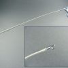
The original Caves-Schulz-Stanford bioptome was modified for left ventricular EMB by reducing the diameter to 6 Fr and increasing the shaft length. This modified Caves-Schulz-Stanford bioptome is frequently used in North America ( Figure 2 ).
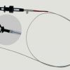
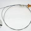
The Cordis bioptome provides smaller myocardial tissue samples compared to those provided by the Caves-Schulz-Stanford bioptome. Today, a modified Cordis bioptome (B-18110; Medizintechnik Meiners, Monheim, Germany) is also used in some European centres [1010. Holzmann M, Nicko A, Kühl U, Noutsias M, Poller W, Hoffmann W, Morguet A, Witzenbichler B, Tschöpe C, Schultheiss HP, Pauschinger M. Holzmann M, Nicko A, Kühl U, Noutsias M, Poller W, Hoffmann W, Morguet A, Witzenbichler B, Tschöpe C, Schultheiss HP, Pauschinger M. Complication rate of right ventricular endomyocardial biopsy via the femoral approach: a retrospective and prospective study analyzing 3048 diagnostic procedures over an 11-year period. Circulation. 2008;118:1722-8.
A comprehensive study evaluating the complication rate of right-ventricular endomyocardial biopsy.] ( Figure 3 ). This bioptome has two jaws with a diameter of 1.8 mm. Compared with the original Cordis bioptome, it has a more flexible polytetrafluoroethylene (Teflon) tube instead of an inflexible steel spiral as well as smaller jaws at the bioptome head.
In numerous German institutions [1111. Yilmaz A, Kindermann I, Kindermann M, Mahfoud F, Ukena C, Athanasiadis A, Hill S, Mahrholdt H, Voehringer M, Schieber M, Klingel K, Kandolf R, Böhm M, Sechtem U. Comparative evaluation of left and right ventricular endomyocardial biopsy: differences in complication rate and diagnostic performance. Circulation. 2010;122:900-9.
A comprehensive study evaluating the risk of complication of left- and right-ventricular endomyocardial biopsy and addressing the issue of CMR-targeted biopsy., 1212. Baccouche H, Mahrholdt H, Meinhardt G, Merher R, Voehringer M, Hill S, Klingel K, Kandolf R, Sechtem U, Yilmaz A. Diagnostic synergy of non-invasive cardiovascular magnetic resonance and invasive endomyocardial biopsy in troponin-positive patients without coronary artery disease. Eur Heart J. 2009;30:2869-79.
A clinical study demonstrating the clinical value of non-invasive CMR and invasive biopsy information and addressing the strengths and limitations of each procedure.], biopsy specimens are taken using either a modified Cordis bioptome or a Maslanka bioptome (EH1518.01-A, H.+H. Maslanka, Tuttlingen, Germany), which are both disposable dedicated cardiac bioptomes ( Figure 4 ). The latter has a pliable shaft and two cutting jaws with a cup diameter of 1.8 mm each. In contrast to the modified Caves-Schulz-Stanford bioptome which has a single cutting jaw and circular cups, the Maslanka bioptome has dual moving jaws and cups with a super-elliptical cutting profile. The shaft of the Maslanka bioptome shows full flexibility over its entire length while the Caves-Schulz-Stanford bioptome is deformable only at the distal curve. The single moving jaw of the Caves-Schulz-Stanford bioptome is actuated by a surgical forceps which is affixed to its proximal end while the Maslanka bioptome is operated by a handle consisting of a thumb ring and slider. There are no major differences between the modified Cordis bioptome and the Maslanka bioptome.
Since the shaft has become smaller in diameter and therefore more flexible, today’s bioptomes are mostly no longer capable of independent movement through the vasculature and therefore have to be positioned with the use of a long guiding sheath or introducing catheter. The guiding sheath itself is introduced into the respective ventricle over a conventional pigtail catheter.
Noteworthy, LV-EMB has also been performed using a “radial” approach and large bore sheathless guiding catheters (e.g. 7.5F EauCath®, ASAHI Intec in combination with a 5.4F Maslanka bioptome) by experienced interventionalists [2626. Schaufele TG, Spittler R, Karagianni A, Ong P, Klingel K, Kandolf R, Mahrholdt H, Sechtem U. Transradial left ventricular endomyocardial biopsy: assessment of safety and efficacy. Clin Res Cardiol. 2015;104(9):773-781. ]. A recent multi-centre study suggested a high success rate of such a radial approach in experienced centres [2727. Choudhury T, Lurz P, Schaufele TG, Menezes MN, Lavi S, Tzemos N, Hartung P, Stiermaier T, Makino K, Bertrand OF, Gilchrist IC, Mamas MA, Bagur R. Radial versus femoral approach for left ventricular endomyocardial biopsy. EuroIntervention. 2019;15(8):678-684. ].
Essential questions regarding the choice of bioptome
- Which ventricle should be biopsied (left vs. right ventricle)?
- Which access site is prefered (jugular vein vs. femoral vein vs. femoral artery vs. radial artery)?
- How many biopsy specimens are required?
A PRACTICAL APPROACH TO LEFT VENTRICULAR ENDOMYOCARDIAL BIOPSY (LV-EMB)
For left ventricular biopsy, heparinisation with unfractionated heparin aiming at an activated clotting time (ACT) target level of 150 sec (which is usually accomplished by a single bolus of 2.500 IU of heparin) is suggested. However, for experienced operators and short procedure times, with careful and repetitive flushing of the guiding sheath (with heparinised sodium chloride solution) after each tissue sampling, intravenous heparinisation is not required.
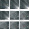
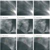
Typically, a long sheath with straight tip is introduced (over a conventional pigtail catheter) through the femoral artery and used for LV-EMB. The tip of the sheath is positioned distal to the mitral apparatus and away from the apex, which has a thinner wall and therefore is more easily perforated than other areas of the left ventricle. The pigtail catheter is then removed, the sheath is flushed, and the respective bioptome is introduced ( Figure 5 ). Alternatively, a coronary guiding catheter which is passed retrogradely through the aortic valve into the left ventricle using a standard J tip guidewire, may be used.
Before insertion, the bioptome is bent smoothly in its distal part to enhance flexibility and to decrease the risk of perforation. Then the bioptome is advanced through the guiding sheath (or guiding catheter) into the left ventricle under biplane fluoroscopic control (30° RAO and 60° LAO or according to the angle of the interventricular septum as determined from the pre-intervention CMR) which helps to guide the tip of the catheter to the target area. Care is taken that the jaws of the bioptome are opened within the left ventricular cavity before close wall contact is reached ( Figure 5 ). Upon reaching wall contact with the opened jaws, pressure is carefully increased as needed to produce a smooth curve at the end of the guiding catheter and the bioptome. Usually close wall contact is accompanied by a series of ventricular premature beats. Then, the biopsy specimen is taken from the wall region of interest. After each biopsy, aspiration of blood from the guiding catheter and rinsing with heparinised sodium chloride solution is performed to prevent clotting. If aspiration is impossible, the sheath needs to be removed and inspected for possible clots, flushed and re-inserted.
A PRACTICAL APPROACH TO RIGHT VENTRICULAR ENDOMYOCARDIAL BIOPSY (RV-EMB)
A long sheath with an angulated tip is introduced through the femoral vein and used for RV-EMB. Alternatively, a coronary guiding catheter which is passed retrogradely through the tricuspide valve into the right ventricle using a standard J tip guidewire, may be used.
The tip of the guiding sheath or guiding catheter is advanced towards the apex of the right ventricle. Next, the tip is gently rotated under fluoroscopic control at 60° LAO or the angle determined from pre-interventional CMR backwards to the spine in order to reach a septal position ( Figure 6 ). Then the bioptome is advanced and the biopsies are taken from the apical septum. Again, care is taken that the jaws of the bioptome are opened within the right ventricular cavity before close wall contact is reached. Upon reaching wall contact with the opened jaws, pressure is carefully increased as needed. At this stage it is recommended that the position of the bioptome is altered once or twice by gently pulling the catheter back with repeated fluoroscopic control of the catheter position both relative to the long-axis of the right ventricle and the interventricular septum. It is important not to take biopsies from a position too close to the pulmonary valve as this may result in chest pain and troponin elevations. Moreover, the right bundle branch may be damaged when attempting to biopsy from such a position.
Essentials in taking biopsy specimens
- Bioptome jaws have to be opened within the respective ventricular cavity “before” myocardial wall contact
- Close wall contact is achieved if a series of ventricular premature beats occurs
- After each biopsy sampling careful and repetitive flushing of the guiding sheath or guiding catheter is mandatory
ALTERNATIVE GUIDING APPROACHES
a) ELECTRO-ANATOMIC MAPPING (EAM)-guided EMB
In case of a patchy disease distribution (e.g. myocarditis or cardiac sarcoidosis), taking EMB samples from the target myocardial area can be challenging. To further optimize the diagnostic yield of EMB in such constellations, some groups have successfully performed electro-anatomic mapping (EAM)-guided EMB sampling [3030. Haanschoten DM, Adiyaman A, 't Hart NA, Jager PL, Elvan A. Value of 3D mapping-guided endomyocardial biopsy in cardiac sarcoidosis: Case series and narrative review on the value of electro-anatomic mapping-guided endomyocardial biopsies. Eur J Clin Invest. 2021;51(4):e13497. ]. Using a high-density mapping catheter, a 3D (bipolar or unipolar) voltage map of the LV and/or RV can be obtained. The respective voltage map (obtained by EAM) allows to differentiate scar tissue from border zones and from viable myocardium based on pre-defined cut-off-values. Thereafter, a bioptome can be connected to the EAM-system and needs to be guided to the target area that is defined by abnormal EAM-based voltage values. Successful applications of this method were shown e.g. in case of suspected cardiac sarcoidosis. However, data on this approach are scarce and it is a quite time-consuming and costly method - and therefore not used for routine work-up.
b) INTRACARDIAC ECHOCARDIOGRAPHY (ICE)-guided BIOPSY
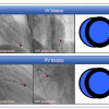
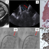
Targeted biopsy of cardiac tumors can be challenging, since precise positioning of the bioptome on the cardiac tumor surface can be tricky due to the glibbery/smooth surface of some tumors with subsequent lateral sliding of the bioptome. In such constellations, intracardiac echocardiography (ICE)-guidance (e.g. with an ACUSON AcuNavTM Ultrasound Catheter, Biosense Webster) of the bioptome to the targeted area can be of paramount value. ICE-guidance may tremendously improve visualization of intracardiac anatomy and thereby provide guidance of the bioptome to the targeted cardiac tumor area. Mostly, ICE-guided biopsy is performed with the additional use of an 8.5-F Agilis NxT steerable sheath/introducer that allows to locate the tip of the bioptome at the target area (visualized by ICE) [3131. Pearman JL, Wall SL, Chen L, Rogers JH. Intracardiac echocardiographic-guided right-sided cardiac biopsy: Case series and literature review. Catheter Cardiovasc Interv. 2020. ]. An example of an ICE-guided biopsy of a cardiac tumor is shown in the Figure 7 (and corresponding Movie 1 and Movie 2). ICE-guidance may play an increasing role for targeted biopsies in the future, but so far available data are limited.
POST-INTERVENTION PATIENT SURVEILLANCE AND CONTROL STUDIES
Immediately after the EMB procedure, echocardiography should be repeated in case of symptoms or systematically within 24 hours in every patient in order to assess whether a new or larger pericardial effusion is present following biopsy. ECG- and blood pressure-monitoring should be continued for at least 12 hours after the procedure.
Potential peri- and post-intervention complications
The feasibility and safety of a medical procedure depends – apart from the complexity of the procedure itself and the clinical status of the patient - primarily on the expertise of the institution or operator. Reported data on the complication risks of EMBs are exclusively based on single-centre experiences. In principal, potential major complications comprise of pericardial tamponade with need for perciardiocentesis, haemo- and pneumopericardium, permanent AV-block requiring permanent pacemaker implantation (especially in patients with pre-existing bundle branch block), myocardial infarction, transient cerebral ischaemic attack and stroke, severe valvular damage and death. Minor complications comprise transient chest pain, ECG changes, arrhythmias, hypotension, small pericardial effusions and complications at the vascular access site.
The overall complication rate varies between <1% and up to 6% for individual centres taking EMBs mainly from the right ventricular septum including sometimes large numbers of repetitive procedures in patients following heart transplantation [1313. Deckers JW, Hare JM, Baughman KL. Complications of transvenous right ventricular endomyocardial biopsy in adult patients with cardiomyopathy: a seven-year survey of 546 consecutive diagnostic procedures in a tertiary referral center. J Am Coll Cardiol. 1992;19:43-7. , 1414. Fowles RE, Mason JW. Endomyocardial biopsy. Ann Intern Med. 1982;97:885-94.
A historical review addressing technical and diagnostic issues regarding endomyocardial biopsy., 1515. Mason JW. Techniques for right and left ventricular endomyocardial biopsy. Am J Cardiol. 1978;41:887-92. , 1616. Starling RC, Van Fossen DB, Hammer DF Unverferth DV. Morbidity of endomyocardial biopsy in cardiomyopathy. Am J Cardiol. 1991;68:133-6. ]. In these studies, the complication rate associated directly with the biopsy procedure itself varied between 0.28% and 3.3%. The largest series reported today by Holzmann et al comprised 3,048 RV-EMB procedures. They found major complications in only 0.12% in the retrospective part of their study and 0% in the prospective part while minor complications were observed in 0.20%/5.5% (retrospective/prospective study part) of patients [1010. Holzmann M, Nicko A, Kühl U, Noutsias M, Poller W, Hoffmann W, Morguet A, Witzenbichler B, Tschöpe C, Schultheiss HP, Pauschinger M. Holzmann M, Nicko A, Kühl U, Noutsias M, Poller W, Hoffmann W, Morguet A, Witzenbichler B, Tschöpe C, Schultheiss HP, Pauschinger M. Complication rate of right ventricular endomyocardial biopsy via the femoral approach: a retrospective and prospective study analyzing 3048 diagnostic procedures over an 11-year period. Circulation. 2008;118:1722-8.
A comprehensive study evaluating the complication rate of right-ventricular endomyocardial biopsy.].
When the study of Holzmann et al is excluded, complication rates published for LV-EMB were considerably lower than for RV-EMB: e.g., Mason [1515. Mason JW. Techniques for right and left ventricular endomyocardial biopsy. Am J Cardiol. 1978;41:887-92. ] performed 161 LV-EMBs and Frustaci et al [1717. Frustaci A, Pieroni M, Chimenti C. The role of endomyocardial biopsy in the diagnosis of cardiomyopathies. Ital Heart J. 2002;3:348-53.
An interesting review addressing the historical development and clinical value of endomyocardial biopsy.] took 1144 LV-EMBs and both documented a major complication rate of 0% for LV-EMB. The results of a comprehensive analysis which directly compared the safety of both procedures (LV-EMB and RV-EMB) in the same patients are in line with these aforementioned studies [1111. Yilmaz A, Kindermann I, Kindermann M, Mahfoud F, Ukena C, Athanasiadis A, Hill S, Mahrholdt H, Voehringer M, Schieber M, Klingel K, Kandolf R, Böhm M, Sechtem U. Comparative evaluation of left and right ventricular endomyocardial biopsy: differences in complication rate and diagnostic performance. Circulation. 2010;122:900-9.
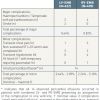
A comprehensive study evaluating the risk of complication of left- and right-ventricular endomyocardial biopsy and addressing the issue of CMR-targeted biopsy.]. Within a study group of 755 patients, a total of 6,361 biopsy samples were taken. LV-EMB was performed in 622 and RV-EMB in 490 patients. During LV-EMB four major complications (two perforations and two peri-/post-interventional strokes) were observed whereas during RV-EMB four perforations leading to haemopericardium and pericardial tamponade with need for pericardiocentesis occurred ( Table 2).Thus, the major complication rate for LV-EMB was 0.64% and for RV-EMB 0.82%. Observed minor complications comprised of transient chest pain (1 case following LV-EMB and 3 following RV-EMB), non-sustained VTs (defined as ≥10 ventricular complexes; 3 cases following LV-EMB and 3 following RV-EMB), transient hypotension (4 cases following RV-EMB) and an AV-block III° requiring a temporary pacemaker in one patient during RV-EMB ( Table 2). Thus, depending on the assignment of those pericardial effusions (to RV- or LV-EMB), the minor complication rate for LV-EMB was either 0.64% (minimum) or 2.89% (maximum) whereas this rate for RV-EMB was either 2.24% (minimum) or 5.10% (maximum). Taken together, both LV- and RV-EMB are safe procedures, with LV-EMB being slightly safer than RV-EMB with respect to the frequency of minor complications.
However, it needs to be considered that the EMB’s complication rate is also dependent on the underlying cardiac disease: In this context, a recent multi-centre study showed an EMB complication rate of 3.13% in patients with cardiac amyloidosis [2828. Chatzantonis G, Bietenbeck M, Elsanhoury A, Tschope C, Pieske B, Tauscher G, Vietheer J, Shomanova Z, Mahrholdt H, Rolf A, Kelle S, Yilmaz A. Diagnostic value of cardiovascular magnetic resonance in comparison to endomyocardial biopsy in cardiac amyloidosis: a multi-centre study. Clin Res Cardiol. 2021;110(4):555-568. ]. Those data indicate a more “fragile” myocardium in case of cardiac amyloidosis with a higher risk of myocardial perforation in particular in case of RV-EMB. Hence, LV-EMB should be the preferred approach in case of suspected cardiac amyloidosis if the respective expertise is available.
As outlined recently [2828. Chatzantonis G, Bietenbeck M, Elsanhoury A, Tschope C, Pieske B, Tauscher G, Vietheer J, Shomanova Z, Mahrholdt H, Rolf A, Kelle S, Yilmaz A. Diagnostic value of cardiovascular magnetic resonance in comparison to endomyocardial biopsy in cardiac amyloidosis: a multi-centre study. Clin Res Cardiol. 2021;110(4):555-568. ], it should be emphasized that the risk of perforation in case of EMB is driven by (a) the properties of the bioptome, (b) the experience of the interventionalist and (c) the composition of the myocardium - with a potentially more “fragile” myocardium in case of CA.
Potential major complications of endomyocardial biopsy
- Pericardial tamponade with need for perciardiocentesis
- Haemo- and pneumopericardium
- Permanent AV-block requiring permanent pacemaker implantation
- Myocardial infarction
- Transient cerebral ischaemic attack and stroke
- Death
Required analyses of EMB samples
According to the recommendations of the American Heart Association (AHA)/American College of Cardiology (ACC)/European Societey of Cardiology (ESC), biopsy samples should be obtained from >1 region per ventricle and the number of samples should range from five to 10 per ventricle, depending on the histopathological studies to be performed [99. Cooper LT, Baughman KL, Feldman AM, Frustaci A, Jessup M, Kuhl U, Levine GN, Narula J, Starling RC, Towbin J, Virmani R. The role of endomyocardial biopsy in the management of cardiovascular disease: a scientific statement from the American Heart Association, the American College of Cardiology, and the European Society of Cardiology. Endorsed by the Heart Failure Society of America and the Heart Failure Association of the European Society of Cardiology. Eur Heart J. 2007;28:3076-93.
Clear scientific statements regarding the appropriate indications for endomyocardial biopsy.]. By selecting >1 region and sampling ≥5 specimens per ventricle, the potential “sampling error” (the likelihood of obtaining false negative results) is minimised [1818. Hauck AJ, Kearney DL, Edwards WD. Evaluation of postmortem endomyocardial biopsy specimens from 38 patients with lymphocytic myocarditis: implications for role of sampling error. Mayo Clin Proc. 1989;64:1235-45.
Interesting study addressing an important limitation of endomyocardial biopsy: sampling error.]. Careful handling of the specimens is required, since artefacts may easily occur and hamper subsequent correct specimen analysis. Therefore, a sterile needle should be carefully used in order to transfer the specimen from the biopsy jaws to the fixative. For subsequent light microscopic or immunohistochemical analyses, the obtained specimens should be fixed in buffered 4% formaldehyde solution for paraffin-embedding which should be stored at room temperature. If transmission electron microscopic examination is required or intended, then at least one to two specimens (per ventricle) should be fixed in 2% glutaraldehyde solution (again at room temperature). If polymerase chain reaction (PCR) is intended for the detection of potential viral genomes, then special RNAlater-solution must be used as appropriate fixative (again at room temperature). If specific immunohistochemistry and/or enzymatic studies are aimed at (e.g. respiratory chain oxidative metabolism in suspected mitochondriopathy), then snap freezing using liquid nitrogen may be appropriate.
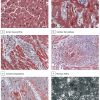
A suggested standard histopathological work-up comprises staining with haematoxylin/eosin, Masson’s trichrome as well as Giemsa and immunohistochemistry using specific antibodies for detection of e.g. CD3+ T-lymphocytes and e.g., CD68+ macrophages. However, depending on the patient’s history and the clinical suspicion and indication for performing EMB, additional analyses such as specific immunohistochemistry, congo-red-staining, amyloid-staining, dystrophin-staining and/or electron microscopy need to be performed ( Figure 8 ).
In addition, in case of suspected myocarditis nested polymerase chain reaction (PCR)/reverse transcriptase–polymerase chain reaction can be performed using samples stored in the aforementioned RNAlater-solution for the detection of e.g. enteroviruses (including coxsackieviruses of group B, various coxsackieviruses of group A and echoviruses), parvovirus B19 (PVB19), adenoviruses, human cytomegalovirus, Epstein-Barr virus, and human herpes virus type 6 (HHV6).
Suggested routine work-up of EMB samples
- Histopathological work-up comprising hematoxylin/eosin, Masson’s trichrome as well as Giemsa stains
- Immunohistochemistry using specific antibodies for detection of T-lymphocytes (e.g., CD3+) and macrophages (e.g., CD68+)
- Molecular pathological PCR analyses for detection of potential viral genomes
Limitations of endomyocardial biopsy
Although both LV- and RV-EMB are safe procedures, both have a non-negligible complication rate. In principle, one would prefer to biopsy just one ventricle if the diagnostic yield was sufficient and omitting biopsy of the other ventricle would not result in missing diagnoses. However, recent data suggest that significantly more diagnostic EMB results are obtained in those patients who undergo biventricular EMBs as compared to those who undergo only selective LV-EMB or selective RV-EMB – at least in case of suspected myocarditis [1111. Yilmaz A, Kindermann I, Kindermann M, Mahfoud F, Ukena C, Athanasiadis A, Hill S, Mahrholdt H, Voehringer M, Schieber M, Klingel K, Kandolf R, Böhm M, Sechtem U. Comparative evaluation of left and right ventricular endomyocardial biopsy: differences in complication rate and diagnostic performance. Circulation. 2010;122:900-9.
A comprehensive study evaluating the risk of complication of left- and right-ventricular endomyocardial biopsy and addressing the issue of CMR-targeted biopsy.]. This finding may be due to a significantly higher number of biopsy samples taken in patients undergoing biventricular EMBs and thereby increasing the diagnostic yield of the procedure, but it is currently not known whether increasing the number of biopsies from one ventricle will result in a similar high diagnostic yield.
Previous data show that in patients who underwent biventricular biopsy for work-up of non-ischaemic cardiomyopathy and/or suspicion of myocarditis (with a similar number of samples taken from both ventricles), significantly more myocardial inflammation was detected than biopsies from a single ventricle [1111. Yilmaz A, Kindermann I, Kindermann M, Mahfoud F, Ukena C, Athanasiadis A, Hill S, Mahrholdt H, Voehringer M, Schieber M, Klingel K, Kandolf R, Böhm M, Sechtem U. Comparative evaluation of left and right ventricular endomyocardial biopsy: differences in complication rate and diagnostic performance. Circulation. 2010;122:900-9.
A comprehensive study evaluating the risk of complication of left- and right-ventricular endomyocardial biopsy and addressing the issue of CMR-targeted biopsy.]: Omitting the LV-EMB resulted in missing 18.7% of cases with myocardial inflammation whereas leaving out the RV-EMB missed only 7.9% of patients with inflammatory disease (p=0.002). Considering the therapeutic and prognostic relevance of myocardial inflammation in EMBs as previously demonstrated by Frustaci et al [1919. Frustaci A, Russo MA, Chimenti C. Randomized study on the efficacy of immunosuppressive therapy in patients with virus-negative inflammatory cardiomyopathy: the TIMIC study. Eur Heart J. 2009;30:1995-2002
A prospective, randomised, placebo-controlled trial demonstrating the importance of biopsy information and the efficacy of immunosuppressive therapy in patients with virus-negative inflammatory cardiomyopathy.] and Kindermann et al [2020. Kindermann I, Kindermann M, Kandolf R Klingel K, Bültmann B, Müller T, Lindinger A, Böhm M. Predictors of outcome in patients with suspected myocarditis. Circulation. 2008;118:639-48. ], valuable diagnostic EMB data will be missed if biopsies are taken only from one ventricle and particularly if only taken from the right ventricle. Therefore, regarding work-up of suspected myocarditis, it is suggested that operators firstly, perform biventricular biopsies to maximise the diagnostic yield as compared to selective biopsies and secondly, perform preferentially LV-EMB if minimising the length of the procedure is a consideration since LV-EMB gives a higher diagnostic yield as compared to RV-EMB while having a lower risk for minor complications.
In recent years, CMR has been established as a non-invasive alternative to diagnosing myocarditis and other forms of cardiomyopathies appropriately [2525. Ferreira VM, Schulz-Menger J, Holmvang G, Kramer CM, Carbone I, Sechtem U, Kindermann I, Gutberlet M, Cooper LT, Liu P, Friedrich MG. Cardiovascular Magnetic Resonance in Nonischemic Myocardial Inflammation: Expert Recommendations. J Am Coll Cardiol. 2018;72(24):3158-3176. , 2828. Chatzantonis G, Bietenbeck M, Elsanhoury A, Tschope C, Pieske B, Tauscher G, Vietheer J, Shomanova Z, Mahrholdt H, Rolf A, Kelle S, Yilmaz A. Diagnostic value of cardiovascular magnetic resonance in comparison to endomyocardial biopsy in cardiac amyloidosis: a multi-centre study. Clin Res Cardiol. 2021;110(4):555-568. ]. The diagnostic performance of both CMR and endomyocardial biopsy was previously evaluated in the same patients with clinically suspected myocarditis [1212. Baccouche H, Mahrholdt H, Meinhardt G, Merher R, Voehringer M, Hill S, Klingel K, Kandolf R, Sechtem U, Yilmaz A. Diagnostic synergy of non-invasive cardiovascular magnetic resonance and invasive endomyocardial biopsy in troponin-positive patients without coronary artery disease. Eur Heart J. 2009;30:2869-79.
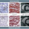
A clinical study demonstrating the clinical value of non-invasive CMR and invasive biopsy information and addressing the strengths and limitations of each procedure.]: EMB results were found to be superior to contrast-enhanced CMR-imaging in diagnosing myocarditis, because of its capabilities based on immunohistochemistry and nested-PCR enabling the detection of even minor forms such as borderline myocarditis, or virus genome presence in the absence of severe myocardial inflammation. Contrast-enhanced CMR-imaging often missed these more subtle forms of myocarditis, possibly due to the limited spatial resolution ( Figure 9). Thus, the value of contrast-enhanced CMR imaging for successful diagnosis of myocarditis seems to be related to the histological degree and extent of inflammation. Nevertheless, it seems reasonable to initially perform CMR in patients with clinically suspected myocarditis and/or non-ischaemic cardiomyopathy, since novel CMR techniques (such as T1- and T2-mappign) promise a higher diagnostic yield [2525. Ferreira VM, Schulz-Menger J, Holmvang G, Kramer CM, Carbone I, Sechtem U, Kindermann I, Gutberlet M, Cooper LT, Liu P, Friedrich MG. Cardiovascular Magnetic Resonance in Nonischemic Myocardial Inflammation: Expert Recommendations. J Am Coll Cardiol. 2018;72(24):3158-3176. ] and since there is a high diagnostic conformity between CMR-based and biopsy-based results [1212. Baccouche H, Mahrholdt H, Meinhardt G, Merher R, Voehringer M, Hill S, Klingel K, Kandolf R, Sechtem U, Yilmaz A. Diagnostic synergy of non-invasive cardiovascular magnetic resonance and invasive endomyocardial biopsy in troponin-positive patients without coronary artery disease. Eur Heart J. 2009;30:2869-79.
A clinical study demonstrating the clinical value of non-invasive CMR and invasive biopsy information and addressing the strengths and limitations of each procedure.].
If the initial CMR study establishes the diagnosis, additional biopsy is unlikely to change this diagnosis [1111. Yilmaz A, Kindermann I, Kindermann M, Mahfoud F, Ukena C, Athanasiadis A, Hill S, Mahrholdt H, Voehringer M, Schieber M, Klingel K, Kandolf R, Böhm M, Sechtem U. Comparative evaluation of left and right ventricular endomyocardial biopsy: differences in complication rate and diagnostic performance. Circulation. 2010;122:900-9.
A comprehensive study evaluating the risk of complication of left- and right-ventricular endomyocardial biopsy and addressing the issue of CMR-targeted biopsy., 1212. Baccouche H, Mahrholdt H, Meinhardt G, Merher R, Voehringer M, Hill S, Klingel K, Kandolf R, Sechtem U, Yilmaz A. Diagnostic synergy of non-invasive cardiovascular magnetic resonance and invasive endomyocardial biopsy in troponin-positive patients without coronary artery disease. Eur Heart J. 2009;30:2869-79.
A clinical study demonstrating the clinical value of non-invasive CMR and invasive biopsy information and addressing the strengths and limitations of each procedure., 2828. Chatzantonis G, Bietenbeck M, Elsanhoury A, Tschope C, Pieske B, Tauscher G, Vietheer J, Shomanova Z, Mahrholdt H, Rolf A, Kelle S, Yilmaz A. Diagnostic value of cardiovascular magnetic resonance in comparison to endomyocardial biopsy in cardiac amyloidosis: a multi-centre study. Clin Res Cardiol. 2021;110(4):555-568. ]. However, in the case of an initial CMR study that is non-conclusive but a diagnosis still needed, for instance in a patient with persistent symptoms suggestive of myocarditis or clinical suspicion of cardiac amyloidosis, biopsy can be employed as a second step. Using this strategy, biopsy may potentially capture e.g. more subtle forms of myocarditis or specific subtypes of cardiac amyloidosis, which cannot be detected with CMR alone. However, although such an algorithm will avoid biopsies in a substantial number of patients - minimising the risk of complications associated with just obtaining a diagnosis, it is obviously not a perfect alternative approach. If the diagnosis of myocarditis and/or cardiomyopathy is merely based on the CMR study, then less detailed information about the degree of inflammation, the presence of special forms of myocarditis (such as giant cell or eosinophilic myocarditis which require specific therapies), or the presence and type of virus is available.
Finally, the tremendous “diagnostic progress” that was achieved by different non-invasive imaging modalities in the last years needs to be carefully considered prior to indicating the performance of invasive EMB. For example, both bone scintigraphy and CMR allow an accurate non-biopsy diagnosis of cardiac transthyretin-amyloidosis (ATTR-CA) when combined with monoclonal protein studies [2828. Chatzantonis G, Bietenbeck M, Elsanhoury A, Tschope C, Pieske B, Tauscher G, Vietheer J, Shomanova Z, Mahrholdt H, Rolf A, Kelle S, Yilmaz A. Diagnostic value of cardiovascular magnetic resonance in comparison to endomyocardial biopsy in cardiac amyloidosis: a multi-centre study. Clin Res Cardiol. 2021;110(4):555-568. , 2929. Gillmore JD, Maurer MS, Falk RH, Merlini G, Damy T, Dispenzieri A, Wechalekar AD, Berk JL, Quarta CC, Grogan M, Lachmann HJ, Bokhari S, Castano A, Dorbala S, Johnson GB, Glaudemans AW, Rezk T, Fontana M, Palladini G, Milani P, Guidalotti PL, Flatman K, Lane T, Vonberg FW, Whelan CJ, Moon JC, Ruberg FL, Miller EJ, Hutt DF, Hazenberg BP, Rapezzi C, Hawkins PN. Nonbiopsy Diagnosis of Cardiac Transthyretin Amyloidosis. Circulation. 2016;133(24):2404-2412. ]. However, in addition to non-invasive imaging data, we still need the histopathological information from EMBs in order to better understand our non-invasive imaging findings - as outlined recently [2525. Ferreira VM, Schulz-Menger J, Holmvang G, Kramer CM, Carbone I, Sechtem U, Kindermann I, Gutberlet M, Cooper LT, Liu P, Friedrich MG. Cardiovascular Magnetic Resonance in Nonischemic Myocardial Inflammation: Expert Recommendations. J Am Coll Cardiol. 2018;72(24):3158-3176. ]: Considering upcoming specific therapies to treat e.g. transthyretin-associated cardiac amyloidosis (ATTR-CA), we will need the EMB information in order to better understand both (a) the therapeutic effect of the respective medication/therapy and (b) the change in non-invasive cardiac imaging parameters that will be used for disease monitoring.
Limitations of endomyocardial biopsy
- Invasive procedure with an albeit low risk of complication
- Limited steerability of bioptomes preventing biopsy sampling in some areas
- Sampling error due to limited number of biopsy samples and limitation of sampling to subendocardial layers
Conclusions
Despite tremendous advances in the field of non-invasive cardiac imaging allowing improved diagnosis of both inflammatory and non-inflammatory (e.g. infiltrative) cardiomyopathies, careful analysis of EMB samples in experienced centres remains the standard of reference for the diagnosis of the majority of cardiac diseases. When performed in high volume centres by experienced interventionalists, EMB has a very low complication rate even when samples are obtained from both ventricles. Considering the important therapeutic consequences of obtaining the correct diagnosis, the risk appears justified in most cases – following an appropriate and careful individual assessment. Similar to other cardiac interventions, it is critical to select the appropriate patient who may benefit from the results of EMB.
Personal perspective – Ali Yilmaz
In spite of great ongoing advances in non-invasive cardiovascular imaging modalities such as computed tomography (CT), cardiovascular magnetic resonance (CMR), positron emission tomography (PET) or their combined use as hybrid techniques in the last years, invasive endomyocardial biopsy (EMB) will continue to be the gold-standard for definitive diagnoses in patients with unexplained cardiomyopathy in the near future due to several reasons:
1) Histopathological EMB analyses comprising immunohistochemical and electron microscopic studies still have an unsurpassed (subcellular) resolution capacity compared to non-invasive imaging modalities;
2) Sophisticated histopathological EMB analysis methods enable detailed diagnoses with unreached diagnostic specificity compared to non-invasive imaging modalities;
3) Essential technical advances have turned EMB into a very safe procedure with a low complication risk thereby further justifying its increased use in appropriate clinical indications. Novel methods for EMB-guidance are either already available (such as ICE-guided biopsy) or not far away from clinical use (such as CMR-based tracking/navigation of the bioptome) and will further stimulate the use of targeted EMBs.
However, the need for additional invasive EMB will decrease in case of some specific cardiomyopathies – such as cardiac amyloidosis or cardiac sarcoidosis – since non-invasive imaging methods such as CMR-based mapping techniques promise a high-yield, comprehensive and easily repeatable diagnostic method - without being prone to a sampling error.
Nevertheless, we still need the histopathological information from EMBs in order to better understand our non-invasive imaging findings e.g. regarding the change in non-invasive cardiac imaging parameters that are used for disease monitoring.
Hence, we believe EMB will continue to be an essential diagnostic procedure in the armamentarium of cardiac interventionalists, however, combined with novel approaches regarding e.g. navigation of the bioptome.


